Tuesday, 21 June 2011


Monday, 20 June 2011
responses to human anatomy post
1.One of the best treatments for acid reflux is dietary modification. Along with the remediesmentioned in this post, you can also try eating small, frequent meals; eat foods that are high in carbohydrates, avoid high-fat meals and limit alcohol.
Rudy Shaw from IBS Diet recently posted..IBS diet – What is right for me 2.Always monitor your food intake and develop a healthy eating habit so you can avoid gaining excess weight
Rudy Shaw from IBS Diet recently posted..IBS diet – What is right for me
2.Always monitor your food intake and develop a healthy eating habit so you can avoid gaining excess weight3.One of the best ways to help acid reflux is to watch your diet. Eat 7-8 small meals instead of 2 or 3 big ones.
Heartburn Treatment recently posted..Heartburn and Acid Reflux Treatments Cures Remedies4.The abdominal area is more important than belly fat of fitness for muscle mass. It is the factory of the body and our overall health mainly depends of this area.
Heartburn Treatment recently posted..Heartburn and Acid Reflux Treatments Cures Remedies
4.The abdominal area is more important than belly fat of fitness for muscle mass. It is the factory of the body and our overall health mainly depends of this area.human anatomy - abdomen
Filed under: Colon Health,Conditions & Diseases,Digestive System
- Human Anatomy – Teeth
- Human Anatomy – Tonsils
- Human Anatomy – Tongue
- Human Anatomy – Esophagus
- Human Anatomy – Stomach
- Human Anatomy – Liver
- Human Anatomy – Gallbladder
- Human Anatomy – Pancreas
- Human Anatomy – Spleen
- Human Anatomy – Appendix
- Human Anatomy – Intestines
- Human Anatomy – Colon
- Human Anatomy – Abdomen
- Human Anatomy – Bladder
- Human Anatomy – Kidneys
- Human Anatomy – Prostate
- Human Anatomy – Penis
- Human Anatomy – Vagina
- Human Anatomy – Heart
- Human Anatomy – Aorta
- Human Anatomy – Skin
- Human Anatomy – Thyroid
- Human Anatomy – Lungs
- Human Anatomy – Brain
- Human Anatomy – Eyes
- Human Anatomy – Ears
- Human Anatomy – Sinuses
- Human Anatomy – Trachea
- Human Anatomy – Blood
- Human Anatomy – Rotator Cuff
- Human Anatomy – Shoulder
- Human Anatomy – Feet
- Human Anatomy – Hair
- Human Anatomy – Achilles Tendon
Besides skin, the abdomen is protected in the front by a thin crust of tissue known as the fascia. The flipside of the abdomen is covered with back muscles.
Several vital blood vessels ply through the abdomen, such as the aorta and the inferior vena cava.

Abdomen conditions and diseases
At times, the abdomen may protrude due to an upsurge of fluids therein, a condition called ascites. Similarly, the abdomen may swell (abdominal distension) because of an increase of gas in the intestines.These and other abdominal complaints are likely to be signs of many underlying conditions, because the abdomen holds a plethora of organs. For example, peritonitis, known as inflammation of the peritoneum or “acute abdomen,” is usually due to an infected or ruptured organ in the abdomen.
Here are some abdominal diseases and conditions:
- Hepatitis. Liver inflammation, often caused by viruses, certain medicines and a weak immune system
- Cirrhosis. Due to chronic inflammation caused by alcoholism and chronic hepatitis, the liver gets scarred.
- Pancreatitis. Pancrea inflammation. Alcohol and gallstones usually cause this condition.
- Peptic ulcer disease. High acidity in the stomach or duodenum (a part of the small intestine) causes erosions or ulcers in the linings of these organs. Causes include aHelicobacter pylori infection or reaction to anti-inflammatory drugs, e.g. ibuprofen.
- Appendicitis. Appendix, an organ in the lower large intestine, gets inflamed. Surgical removal is usually recommended for an inflamed appendix.
- Gastritis. Stomach inflammation, oft-caused by alcohol, NSAIDs and bacterial infection
- Cholecystitis. Gallbladder inflammation, caused by a gallstone obstructing a gallbladder duct
- Aortic aneurysm. Occurs when the wall of the aorta in the abdomen weakens. The aorta then expands like a balloon over the years until it bursts.
- Abdominal hernia. Weakening of the fascia
- Dyspepsia. Another term for a stomach upset/indigestion
- Intestinal obstruction. One part or the entire small/large intestine ceases to function. Abdominal distension and vomiting ensues.
- Gastroparesis. Due to debilitating diseases like diabetes and cancer, the stomach empties at a slower pace than usual, causing vomiting and nausea.
Treating abdomen conditions
Abdominal surgery is always indicated for severe conditions such as colon/stomach cancer, appendicitis, aneurysm and cholecystitis. The surgeon may either choose an open or laparoscopic surgery according to the patient’s case.To treat an overly acid stomach, a doctor prescribes H2 blockers to keep histamine in the body from compelling acid production. In the same way, proton pump inhibitors can hinder the acid-secreting pumps of the stomach.
Other abdomen treatments:
- Antibiotics. Helicobacter pylori, a bacterium notorious for causing many abdominal illnesses, can be successfully countered by prescription antibiotics.
- Motility agents. Gastroparesis and constipation, which are caused by decreased stomach contraction, can be treated with these drugs.
- Laxatives. Used to treat constipation
- DigestAssist – relieves acute and chronic digestive problems.
- Acid Free-Flux – relieves acute acid reflux and heartburn.
- Gastronic Dr – relieves acute and chronic digestive disorders.
- Gasolve Relief – promotes digestive calm and comfort after meals to alleviate gas and bloating.
- Liver Dr – a 100% herbal remedy that supports healthy liver functioning and systemic health.
- Insulate Plus – a 100% herbal remedy that helps maintain healthy blood sugar levels and promotes pancreatic health.
- Diabetonic – a safe, non–addictive, natural remedy containing 100% homeopathic ingredients to balance sugar levels in the blood and pancreas.
- Natural Moves – a 100% natural remedy for constipation.
- ColoFlush – a very effective natural remedy to flush the colon and promote systemic health.
- Bowtrol Probiotic – serve as supplements of good microbes needed by the colon.
Tests for abdomen conditions
Some abdominal conditions are discernible enough without the use of machines. A doctor can just “listen” to your body with a simple tap of the abdomen or a press of the stethoscope.To be sure, the doctor would use tests like an abdominal ultrasound to get detailed images of the abdominal organ in question. If you want to rectify an abdominal condition, resuming a healthy lifestyle thereafter, consider taking these and other abdomen tests:
- Endoscopic retrograde cholangiopancreatography. Used to detect problems in the pancreas. With an endoscope, a doctor puts a tube into a pancreatic duct and squirts a fluid that deflects X-rays.
- Esophagogastroduodenoscopy. Also known as upper endoscopy, this test entails the insertion of an endoscope through the mouth. With a camera on its tip, the endoscope lets the doctor see the stomach and the first parts of the small intestine at magnified range.
- Colonoscopy. Also known as lower endoscopy, this test differs from above in that the endoscope enters the body through the anus, not the mouth. It is mainly used to provide magnified images of the colon and other organs in the lower abdomen.
- pH tests. Monitors acidity in the esophagus. Done by inserting a tube into the nose
- Gastric emptying study. Tests how fast food comes down the stomach. The food is immersed in a radioactive substance, allowing it to “shine” through in the scanner.
- Computed tomography (CT scan). A computer and an X-ray machine work together to provide detailed photos of the abdominal organs.
- Magnetic resonance imaging (MRI scan). Uses radio waves to provide high-resolution pictures of the abdominal organs, especially the pancreas, liver and gallbladder
Sunday, 12 June 2011
Referred pain
If you woke up with a pain in your shoulder, you'd probably think something was wrong with your shoulder, right? Maybe you slept on it the wrong way, maybe you're a weekend warrior who threw the football a few too many times. In most cases, your hunch is probably right. Pain in the shoulder usually indicates an injury or disease that affects a structure in your shoulder, such as, say, your subacromial bursa or a rotator cuff tendon. Makes sense, doesn't it?
But you might be way off. Sometimes the brain gets confused, making you think that one part of the body hurts, when in fact another part of the body, far removed from the pain, is the real source of trouble. This curious (and clinically important) phenomenon is known asreferred pain. For example, it's unlikely but possible that your shoulder pain is a sign of something insidious happening in your liver,gall bladder, stomach, spleen, lungs, or pericardial sac (the connective tissue bag containing the heart). Strange, isn't it? Conditions as diverse as liver abscesses, gallstones, gastric ulcers, splenic rupture, pneumonia, and pericarditis can all cause shoulder pain. How is that possible?
Neuroscientists still don't know precisely which anatomical connections are responsible for referred pain, but the prevailing explanation seems to work pretty well. In a nutshell, referred pain happens when nerve fibers from regions of high sensory input (such as the skin) and nerve fibers from regions of normally low sensory input (such as the internal organs) happen to converge on the same levels of the spinal cord. The best known example is pain experienced during a heart attack. Nerves from damaged heart tissue convey pain signals to spinal cord levels T1-T4 on the left side, which happen to be the same levels that receive sensation from the left side of the chest and part of the left arm. The brain isn't used to receiving such strong signals from the heart, so it interprets them as pain in the chest and left arm.
So what about that shoulder pain? All of organs listed above bump up against the diaphragm, the thin, dome-shaped muscle that moves up and down with every breath. The diaphragm is innervated by twophrenic nerves (left and right), which emerge from spinal cord levelsC3, C4, and C5 (medical students remember these spinal cord levels using the mnemonic, "C3, 4, 5 keeps the diaphragm alive"). The phrenic nerves carry both motor and sensory impulses, so they make the diaphragm move and they convey sensation from the diaphragm to the central nervous system.
Most of the time there isn't any sensation to convey from the diaphragm, at least at the conscious level. But if a nearby organ gets sick, it may irritate the diaphragm, and the sensory fibers of one of the phrenic nerves are flooded with pain signals that travel to the spinal cord (at C3-C5). It turns out that C3 and C4 don't just keep the diaphragm alive; neurons at these two spinal cord levels also receive sensation from the shoulders (via the supraclavicular nerves). So when pain neurons at C3 and C4 sound the alarm, the brain assumes (quite reasonably) that the shoulder is to blame. Usually that's a good assumption, but sometimes it's wrong.
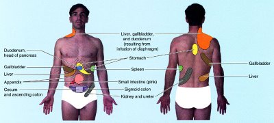 Illustration showing sites of referred pain from abdominal organs. From Moore and Dalley's Clinically Oriented Anatomy. Please note that I added the "tighty whities" with Photoshop (hey, this is a family friendly site).
Another example that seems bizarre until you know the anatomy is disease in the stomach causing pain between the shoulder blades. A classmate told me that one of her college professors complained to his doctor about pain in his upper back that wouldn't go away. It turns out that the professor had gastric cancer, a relatively aggressive and often incurable disease unless it's caught early. Unfortunately it wasn't caught early enough and it ended up taking his life. Maybe the outcome would have been different if the doctor had remembered that some of the nerve fibers to the stomach (specifically, visceral afferents that travel in the greater splanchnic nerve) convey pain signals to the same spinal cord levels (especially T5 and T6) that receive pain signals from the skin between the shoulder blades. This variety of referred pain is rare, but it happens often enough to be mentioned in anatomy textbooks.
Not all cases of referred pain are easy to find in textbooks. Take theappendix, for instance. That wormy little appendage of the colon is usually located in the right lower quadrant of the belly, nowhere near the diaphragm. Typically people with appendicitis feel diffuse pain or discomfort around the belly button, or sharp pain in the right lower quadrant if the appendix is getting ready to burst. But occasionally the pain refers to the right shoulder. Why? Note that I said the appendix is usually located in right lower quadrant. Sometimes, early in embryological development, the colon doesn't rotate as much as it should, and the appendix ends up in the right upper quadrant (or even on the left side of the belly). That malrotation isn't necessarily a problem for the patient until the appendix becomes swollen with infection, irritating both the diaphragm and the doctor who is trying to make a diagnosis!
So if your shoulder happens to hurt right now, should you be worrying about something bad in your belly? Probably not. The most common causes of shoulder pain are, by far, musculoskeletal problems like osteoarthritis, adhesive capsulitis, and rotator cuff tendinitis. Often, with the help of taking it easy and a few rounds of ibuprofen, the pain goes away on its own. But if you're still concerned, make an appointment with your family physician. Red flags include more ominous symptoms like fever, unexplained weight loss, and pain in the left shoulder or scapular area that gets worse with exertion.
Your doctor will want to know all about the pain. Was there a specific injury or did the pain come on slowly? Does the pain occur without movement of the shoulder? Can you make it worse with specific movements? Are there any other symptoms or chronic medical problems? The answers to these questions, along with a physical exam and appropriate imaging studies, will provide the information your doctor needs to understand the problem and create a treatment plan. The odds are good that the problem is relatively common and treatable. But if your doctor seems stumped, it wouldn't hurt to ask, "Is there any chance this could be referred pain?"
But you might be way off. Sometimes the brain gets confused, making you think that one part of the body hurts, when in fact another part of the body, far removed from the pain, is the real source of trouble. This curious (and clinically important) phenomenon is known asreferred pain. For example, it's unlikely but possible that your shoulder pain is a sign of something insidious happening in your liver,gall bladder, stomach, spleen, lungs, or pericardial sac (the connective tissue bag containing the heart). Strange, isn't it? Conditions as diverse as liver abscesses, gallstones, gastric ulcers, splenic rupture, pneumonia, and pericarditis can all cause shoulder pain. How is that possible?
Neuroscientists still don't know precisely which anatomical connections are responsible for referred pain, but the prevailing explanation seems to work pretty well. In a nutshell, referred pain happens when nerve fibers from regions of high sensory input (such as the skin) and nerve fibers from regions of normally low sensory input (such as the internal organs) happen to converge on the same levels of the spinal cord. The best known example is pain experienced during a heart attack. Nerves from damaged heart tissue convey pain signals to spinal cord levels T1-T4 on the left side, which happen to be the same levels that receive sensation from the left side of the chest and part of the left arm. The brain isn't used to receiving such strong signals from the heart, so it interprets them as pain in the chest and left arm.
So what about that shoulder pain? All of organs listed above bump up against the diaphragm, the thin, dome-shaped muscle that moves up and down with every breath. The diaphragm is innervated by twophrenic nerves (left and right), which emerge from spinal cord levelsC3, C4, and C5 (medical students remember these spinal cord levels using the mnemonic, "C3, 4, 5 keeps the diaphragm alive"). The phrenic nerves carry both motor and sensory impulses, so they make the diaphragm move and they convey sensation from the diaphragm to the central nervous system.
Most of the time there isn't any sensation to convey from the diaphragm, at least at the conscious level. But if a nearby organ gets sick, it may irritate the diaphragm, and the sensory fibers of one of the phrenic nerves are flooded with pain signals that travel to the spinal cord (at C3-C5). It turns out that C3 and C4 don't just keep the diaphragm alive; neurons at these two spinal cord levels also receive sensation from the shoulders (via the supraclavicular nerves). So when pain neurons at C3 and C4 sound the alarm, the brain assumes (quite reasonably) that the shoulder is to blame. Usually that's a good assumption, but sometimes it's wrong.
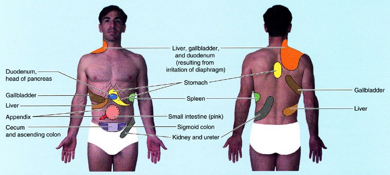 Illustration showing sites of referred pain from abdominal organs. From Moore and Dalley's Clinically Oriented Anatomy. Please note that I added the "tighty whities" with Photoshop (hey, this is a family friendly site).
Another example that seems bizarre until you know the anatomy is disease in the stomach causing pain between the shoulder blades. A classmate told me that one of her college professors complained to his doctor about pain in his upper back that wouldn't go away. It turns out that the professor had gastric cancer, a relatively aggressive and often incurable disease unless it's caught early. Unfortunately it wasn't caught early enough and it ended up taking his life. Maybe the outcome would have been different if the doctor had remembered that some of the nerve fibers to the stomach (specifically, visceral afferents that travel in the greater splanchnic nerve) convey pain signals to the same spinal cord levels (especially T5 and T6) that receive pain signals from the skin between the shoulder blades. This variety of referred pain is rare, but it happens often enough to be mentioned in anatomy textbooks.
Not all cases of referred pain are easy to find in textbooks. Take theappendix, for instance. That wormy little appendage of the colon is usually located in the right lower quadrant of the belly, nowhere near the diaphragm. Typically people with appendicitis feel diffuse pain or discomfort around the belly button, or sharp pain in the right lower quadrant if the appendix is getting ready to burst. But occasionally the pain refers to the right shoulder. Why? Note that I said the appendix is usually located in right lower quadrant. Sometimes, early in embryological development, the colon doesn't rotate as much as it should, and the appendix ends up in the right upper quadrant (or even on the left side of the belly). That malrotation isn't necessarily a problem for the patient until the appendix becomes swollen with infection, irritating both the diaphragm and the doctor who is trying to make a diagnosis!
So if your shoulder happens to hurt right now, should you be worrying about something bad in your belly? Probably not. The most common causes of shoulder pain are, by far, musculoskeletal problems like osteoarthritis, adhesive capsulitis, and rotator cuff tendinitis. Often, with the help of taking it easy and a few rounds of ibuprofen, the pain goes away on its own. But if you're still concerned, make an appointment with your family physician. Red flags include more ominous symptoms like fever, unexplained weight loss, and pain in the left shoulder or scapular area that gets worse with exertion.
Your doctor will want to know all about the pain. Was there a specific injury or did the pain come on slowly? Does the pain occur without movement of the shoulder? Can you make it worse with specific movements? Are there any other symptoms or chronic medical problems? The answers to these questions, along with a physical exam and appropriate imaging studies, will provide the information your doctor needs to understand the problem and create a treatment plan. The odds are good that the problem is relatively common and treatable. But if your doctor seems stumped, it wouldn't hurt to ask, "Is there any chance this could be referred pain?"
the ethics of retail radiology
Hey everyone, I'm currently between graduation and residency, so I've decided to post an essay I wrote for one of the last courses of medical school: Medical Ethics. The essay focuses on a controversial topic in my chosen field, radiology. Enjoy!

From a scientific perspective, however, the effectiveness of whole-body CT as a screening exam is unproven (7,13). Although anecdotal evidence makes the headlines (8), there is "no evidence that total body CT screening is cost efficient or effective in prolonging life" (13). The potential benefit of early detection may be outweighed by adverse effects and wasteful costs (discussed below).
On the other hand, no randomized controlled trial (RCT) - the gold standard for evidence-based medicine - has been designed to study screening with whole-body CT. Supporters argue that the "delay imposed by a randomized clinical trial (perhaps a decade) makes such a trial's results an unacceptable Holy Grail" (9). Other CT screening exams are growing in scientific support as evidence accumulates, at least in certain patient populations (3). Initially controversial, coronary artery calcium screening is now recommended for patients at moderate risk for heart disease (14). CT lung screening in smokers is still intensely debated, but a large RCT - the National Lung Screening Trial - should clarify things (15). CT colonography (virtual colonoscopy) is widely considered an acceptable option for colorectal cancer screening (16). Given the possibly life-saving benefits of a whole-body CT, why should we have to wait for stronger evidence?
Nonmaleficence
Our protocols are built to insure the lowest dose possible, minimizing risk to non-calculable levels. The breast tissue in the chest CT study is exposed to no more radiation than a mammogram (1).
Perhaps the most common ethical dilemmas in clinical medicine involve the balance of beneficence and nonmaleficence, or potential benefit vs. potential harm. Critics claim that the potential benefits of whole-body CT screening exams are exaggerated and the risks are minimized or not discussed at all.
To be embraced by the medical community, screening exams must meet certain criteria. For example, the exam should be good at detecting disease that is both serious and treatable if caught early. Effective screening programs minimize false positives and false negatives and maximize cost effectiveness by focusing on populations in which there is a higher prevalence of the disease. CT colonography, for example, is usually recommended only for patients 50 years or older (16).
Whether whole-body CT could ever meet the criteria of an effective screening program is unknown, but preliminary data are not encouraging. One study based on a hypothetical cohort of 500,000 asymptomatic 50-year-old men found that whole-body CT scans are more expensive, in costs per life-year, than most health care interventions currently funded in U.S.4 One serious concern is the high rate of false positives in populations with a low prevalence of disease. In a recent study of patients seen at one outpatient imaging center in California during a 6-month period, 86% of the 1192 patients (mean age 54 year old, most self-referred) had at least one abnormal finding described in the whole-body screening report (17). In many cases there are no standard criteria for deciding which lesions need further work up. Many findings that do require further evaluation - such as certain renal masses - are likely to be indolent diseases that never would have bothered the patient if they had not been discovered on CT (18).
Additional work up means additional anxiety and risk for the patient. In the California study, 37% of patients received at least one recommendation for further evaluation, most commonly additional imaging of lungs or kidneys with intravenous contrast (17). Indeterminate findings could also lead to invasive procedures such as biopsy, surgery, or endoscopy; however, none of these were specifically recommended in the California study (17).
What about the radiation risk from CT exams? Although the degree of risk has been debated, it appears to be small. One study estimated that the radiation dose from a single whole-body CT screening exam is approximately 15-20 mSv (millisieverts). For a 45-year-old adult receiving a single exam, this dose increases the risk of death from cancer by 0.08% (19). AccuScan claims the dose of its whole-body CT screen is 5.2 mSv, or 1/2 to 1/3 the dose of conventional CT study (1). This dose is comparable to the natural background radiation exposure to the average American in one year (3.1 mSv).
Justice
AccuScan is another tool in the arsenal against disease. It is meant to supplement and enhance traditional health care, not replace it (1).
In bioethics, distributive justice refers primarily to the equitable use of limited health care resources. For critics of retail radiology, the main concern here is the additional demand for medical resources created by false positives and the discovery of indolent disease (17). Consumers pay out-of-pocket for the initial scan, then funnel into the traditional health care system for follow up if recommended. Some physicians may find themselves in the awkward position of having to provide education and management based on a scan they did not order or discuss with the patient before it was performed (6).
Recommendations
In any medical setting, some activities - using fear in advertising, making false promises, failing to provide informed consent - are clearly unethical. Other activities, such as selling CT screening exams directly to the public, are more ethically ambiguous. Precisely where the activities of self-referred imaging centers fall on the ethical continuum depends partly on which principles are most heavily weighted. Proponents tend to emphasize autonomy and beneficence; detractors tend to emphasize nonmaleficence and justice. Here are some recommendations for making the practice of retail radiology as ethical as possible.
References
Image source:
The Ethics of Retail Radiology
Imagine a future where diseases are caught in their infancy and disorders are slowed, stopped and even reversed before patients ever suffer from the symptoms. With AccuScan, that future is available right now, right here in Salt Lake City (1).
Nestled between a megaplex and a boutique in the Gateway outdoor mall, AccuScan Health Imaging is a body imaging center that offers the promise of early disease detection in asymptomatic individuals. No physician referral is required; any customer willing to pay out-of-pocket can choose from a selection of computed tomography (CT) screening exams, including whole body scan, heart scan (coronary artery calcium screen), lung scan, head scan, and virtual colonoscopy. The scans are read by a board-certified radiologist and the patient receives the images, a detailed written report, and referral to a specialist if there are any concerning findings (1).
A decade ago, self-referred body imaging centers were on the rise, fueled by publicity on talk shows, stories in health magazines, direct-to-consumer (DTC) advertising, and dissatisfaction with managed care. Within a few years, however, the retail radiology bubble burst, and many initially successful centers went out of business (2). The collapse has been attributed to economic forces (e.g., competition driving down prices) and the influence of medical professional societies, which have been uniformly skeptical of the value of whole-body screening. Critics have raised many concerns, such as unethical advertising, unnecessary radiation exposure, high false-positive rates, and low cost-effectiveness (3-6). The FDA warns that "whole-body CT screening has not been demonstrated to meet generally accepted criteria for an effective screening procedure" (7).
In spite of these misgivings, the demand for imaging centers like AccuScan has not evaporated completely. They remain unregulated by the FDA (7). A recent survey found that 2% of radiologists still support whole-body CT screening, and many more support CT screening exams of the heart or lungs in particular patients (3). Supporters emphasize the patient’s right to know and argue that the benefits of early disease detection outweigh the risks (8,9).
Are self-referred imaging centers ethical? In this paper I would like to examine the controversy in the light of widely accepted bioethical principles: autonomy, beneficence, nonmaleficence, and justice. How does each principle support or discredit the practices of retail radiology? At the end, rather than simply applying the label of "ethical" or "unethical," I would like to address a more practical question: what can businesses like AccuScan do to function as ethically as possible?
Autonomy
Take control of your own health and stay as healthy as you feel with AccuScan (1).
One of the most compelling arguments in favor of self-referred imaging centers is patient autonomy. Proponents view their services as a form of "medical self-empowerment" (9). Frustrated with a "paternalistic payor-physician culture that controls the practice of medicine and patient referral,"10 some customers are willing to pay more for a different kind of experience. In addition to data the patient cannot obtain from their regular providers, AccuScan promises a visit with "no green scrubs, sterile surroundings, long waits, or impersonal treatment" (1). Patients enjoy the highest possible sense of autonomy because they are paying out-of-pocket for a service of their choice.
However, critics contend that patients are choosing whole-body CT scans with incomplete or even inaccurate information (6). According to the AMA Code of Ethics, the "patient’s right of self-decision can be effectively exercised only if the patient possesses enough information to enable an informed choice" (11). One analysis of print advertisements and brochures concluded that DTC marketing of self-referred imaging services "fails to provide prospective consumers with comprehensive balanced information vital to informed autonomous decision making" (5). For example, there was typically no information on the rate of false-positive findings and the procedures needed to evaluate indeterminate lesions. Many ads conveyed a false sense of reassurance, some evoked fear (5).
Beneficence
Many diseases progress silently, leaving you totally asymptomatic... AccuScan can provide early detection, allowing physicians to slow, stop, or in some cases reverse disease long before your symptoms would have led you to seek help (1).
Most consumers who seek whole-body CT scans and other screening exams are motivated by the benefits of early detection. Indeed, early detection leading to treatment and improved survival rates is the rationale for mammography, colonoscopy, and other scientifically validated screening exams. If screening for disease in selected organs is good, why not go for the whole body? A whole-body CT scan is a rapid (15 minutes), painless, noninvasive method of acquiring an extraordinarily detailed picture of internal anatomy. One radiologist, Richard Penfil, expressed the sentiment this way:
Imagine a future where diseases are caught in their infancy and disorders are slowed, stopped and even reversed before patients ever suffer from the symptoms. With AccuScan, that future is available right now, right here in Salt Lake City (1).
Nestled between a megaplex and a boutique in the Gateway outdoor mall, AccuScan Health Imaging is a body imaging center that offers the promise of early disease detection in asymptomatic individuals. No physician referral is required; any customer willing to pay out-of-pocket can choose from a selection of computed tomography (CT) screening exams, including whole body scan, heart scan (coronary artery calcium screen), lung scan, head scan, and virtual colonoscopy. The scans are read by a board-certified radiologist and the patient receives the images, a detailed written report, and referral to a specialist if there are any concerning findings (1).
A decade ago, self-referred body imaging centers were on the rise, fueled by publicity on talk shows, stories in health magazines, direct-to-consumer (DTC) advertising, and dissatisfaction with managed care. Within a few years, however, the retail radiology bubble burst, and many initially successful centers went out of business (2). The collapse has been attributed to economic forces (e.g., competition driving down prices) and the influence of medical professional societies, which have been uniformly skeptical of the value of whole-body screening. Critics have raised many concerns, such as unethical advertising, unnecessary radiation exposure, high false-positive rates, and low cost-effectiveness (3-6). The FDA warns that "whole-body CT screening has not been demonstrated to meet generally accepted criteria for an effective screening procedure" (7).
In spite of these misgivings, the demand for imaging centers like AccuScan has not evaporated completely. They remain unregulated by the FDA (7). A recent survey found that 2% of radiologists still support whole-body CT screening, and many more support CT screening exams of the heart or lungs in particular patients (3). Supporters emphasize the patient’s right to know and argue that the benefits of early disease detection outweigh the risks (8,9).
Are self-referred imaging centers ethical? In this paper I would like to examine the controversy in the light of widely accepted bioethical principles: autonomy, beneficence, nonmaleficence, and justice. How does each principle support or discredit the practices of retail radiology? At the end, rather than simply applying the label of "ethical" or "unethical," I would like to address a more practical question: what can businesses like AccuScan do to function as ethically as possible?
Autonomy
Take control of your own health and stay as healthy as you feel with AccuScan (1).
One of the most compelling arguments in favor of self-referred imaging centers is patient autonomy. Proponents view their services as a form of "medical self-empowerment" (9). Frustrated with a "paternalistic payor-physician culture that controls the practice of medicine and patient referral,"10 some customers are willing to pay more for a different kind of experience. In addition to data the patient cannot obtain from their regular providers, AccuScan promises a visit with "no green scrubs, sterile surroundings, long waits, or impersonal treatment" (1). Patients enjoy the highest possible sense of autonomy because they are paying out-of-pocket for a service of their choice.
However, critics contend that patients are choosing whole-body CT scans with incomplete or even inaccurate information (6). According to the AMA Code of Ethics, the "patient’s right of self-decision can be effectively exercised only if the patient possesses enough information to enable an informed choice" (11). One analysis of print advertisements and brochures concluded that DTC marketing of self-referred imaging services "fails to provide prospective consumers with comprehensive balanced information vital to informed autonomous decision making" (5). For example, there was typically no information on the rate of false-positive findings and the procedures needed to evaluate indeterminate lesions. Many ads conveyed a false sense of reassurance, some evoked fear (5).
Beneficence
Many diseases progress silently, leaving you totally asymptomatic... AccuScan can provide early detection, allowing physicians to slow, stop, or in some cases reverse disease long before your symptoms would have led you to seek help (1).
Most consumers who seek whole-body CT scans and other screening exams are motivated by the benefits of early detection. Indeed, early detection leading to treatment and improved survival rates is the rationale for mammography, colonoscopy, and other scientifically validated screening exams. If screening for disease in selected organs is good, why not go for the whole body? A whole-body CT scan is a rapid (15 minutes), painless, noninvasive method of acquiring an extraordinarily detailed picture of internal anatomy. One radiologist, Richard Penfil, expressed the sentiment this way:
Historically, you go to your internist once a year and he thumps on your chest, takes out his stethoscope, draws some blood to send to a laboratory, sends you for a chest x-ray, and asks how your golf game is doing. That, to me, is the virtual examination. The actual examination is where you take a look at what is going on inside your body (12).
From a scientific perspective, however, the effectiveness of whole-body CT as a screening exam is unproven (7,13). Although anecdotal evidence makes the headlines (8), there is "no evidence that total body CT screening is cost efficient or effective in prolonging life" (13). The potential benefit of early detection may be outweighed by adverse effects and wasteful costs (discussed below).
On the other hand, no randomized controlled trial (RCT) - the gold standard for evidence-based medicine - has been designed to study screening with whole-body CT. Supporters argue that the "delay imposed by a randomized clinical trial (perhaps a decade) makes such a trial's results an unacceptable Holy Grail" (9). Other CT screening exams are growing in scientific support as evidence accumulates, at least in certain patient populations (3). Initially controversial, coronary artery calcium screening is now recommended for patients at moderate risk for heart disease (14). CT lung screening in smokers is still intensely debated, but a large RCT - the National Lung Screening Trial - should clarify things (15). CT colonography (virtual colonoscopy) is widely considered an acceptable option for colorectal cancer screening (16). Given the possibly life-saving benefits of a whole-body CT, why should we have to wait for stronger evidence?
Nonmaleficence
Our protocols are built to insure the lowest dose possible, minimizing risk to non-calculable levels. The breast tissue in the chest CT study is exposed to no more radiation than a mammogram (1).
Perhaps the most common ethical dilemmas in clinical medicine involve the balance of beneficence and nonmaleficence, or potential benefit vs. potential harm. Critics claim that the potential benefits of whole-body CT screening exams are exaggerated and the risks are minimized or not discussed at all.
To be embraced by the medical community, screening exams must meet certain criteria. For example, the exam should be good at detecting disease that is both serious and treatable if caught early. Effective screening programs minimize false positives and false negatives and maximize cost effectiveness by focusing on populations in which there is a higher prevalence of the disease. CT colonography, for example, is usually recommended only for patients 50 years or older (16).
Whether whole-body CT could ever meet the criteria of an effective screening program is unknown, but preliminary data are not encouraging. One study based on a hypothetical cohort of 500,000 asymptomatic 50-year-old men found that whole-body CT scans are more expensive, in costs per life-year, than most health care interventions currently funded in U.S.4 One serious concern is the high rate of false positives in populations with a low prevalence of disease. In a recent study of patients seen at one outpatient imaging center in California during a 6-month period, 86% of the 1192 patients (mean age 54 year old, most self-referred) had at least one abnormal finding described in the whole-body screening report (17). In many cases there are no standard criteria for deciding which lesions need further work up. Many findings that do require further evaluation - such as certain renal masses - are likely to be indolent diseases that never would have bothered the patient if they had not been discovered on CT (18).
Additional work up means additional anxiety and risk for the patient. In the California study, 37% of patients received at least one recommendation for further evaluation, most commonly additional imaging of lungs or kidneys with intravenous contrast (17). Indeterminate findings could also lead to invasive procedures such as biopsy, surgery, or endoscopy; however, none of these were specifically recommended in the California study (17).
What about the radiation risk from CT exams? Although the degree of risk has been debated, it appears to be small. One study estimated that the radiation dose from a single whole-body CT screening exam is approximately 15-20 mSv (millisieverts). For a 45-year-old adult receiving a single exam, this dose increases the risk of death from cancer by 0.08% (19). AccuScan claims the dose of its whole-body CT screen is 5.2 mSv, or 1/2 to 1/3 the dose of conventional CT study (1). This dose is comparable to the natural background radiation exposure to the average American in one year (3.1 mSv).
Justice
AccuScan is another tool in the arsenal against disease. It is meant to supplement and enhance traditional health care, not replace it (1).
In bioethics, distributive justice refers primarily to the equitable use of limited health care resources. For critics of retail radiology, the main concern here is the additional demand for medical resources created by false positives and the discovery of indolent disease (17). Consumers pay out-of-pocket for the initial scan, then funnel into the traditional health care system for follow up if recommended. Some physicians may find themselves in the awkward position of having to provide education and management based on a scan they did not order or discuss with the patient before it was performed (6).
Recommendations
In any medical setting, some activities - using fear in advertising, making false promises, failing to provide informed consent - are clearly unethical. Other activities, such as selling CT screening exams directly to the public, are more ethically ambiguous. Precisely where the activities of self-referred imaging centers fall on the ethical continuum depends partly on which principles are most heavily weighted. Proponents tend to emphasize autonomy and beneficence; detractors tend to emphasize nonmaleficence and justice. Here are some recommendations for making the practice of retail radiology as ethical as possible.
- Perhaps most importantly, providers of any screening exam must obtain true informed consent (20). Patients do have a right to make decisions about their own health care, even if the physician does not agree with the decision. However, respect for autonomy does not mean the patient should make health care decisions in a vacuum. An important responsibility of the physician is to create the conditions for a truly informed choice about procedures and tests, making sure the patient understands the benefits, risks, and limitations. Patients are often aware of a small radiation risk, but are less likely to understand the risks of false positives and the potentially invasive and costly work up and treatment of indolent disease. Patients should be warned against a false sense of security that comes with a negative exam.
- Advertisements should be free of unsubstantiated scientific claims, exaggerated promises, or scare tactics. If abuses continue, DTC marketing of imaging services should be subjected to regulatory control similar to what currently exists for the pharmaceutical industry (7).
- Providers should be wary of screening everyone who walks in the door. Patients should be informed of the evidence-based guidelines, if any, for the screening exams they would like to receive. Of course, a challenge for all medical entrepreneurs is putting the patient's welfare ahead of financial interests. According to an early advocate of CT screening, "most centers attempt to exclude individuals who are younger than 40 years old, but the financial incentive clearly tempts some to lower the threshold" (9).
These recommendations will not satisfy everyone, but I believe they are a step in the right direction. Whether retail radiology could follow them and stay in business is an open question.
References
- AccuScan Health Imaging. Available athttp://www.accuscanhealthimaging.com/
- Kolata G. Rapid rise and fall for body-scanning clinics. New York Times, 23 Jan 2005. Available athttp://www.nytimes.com/
- Burger IM, Kass NE, Sunshine JH, Siegelman SS. The use of CT for screening: A national survey of radiologists' activities and attitudes. Radiology 2008; 248:160-168.
- Beinfeld MT, Wittenberg E, Gazelle GS. Cost-effectiveness of whole-body CT screening. Radiology 2005; 234:415-422.
- Illes J, Kann D, Karetsky K, et al. Advertising, patient decision making, and self-referral for computed tomographic and magnetic resonance imaging. Arch Intern Med 2004; 164:2415-2419.
- O'Malley PG, Taylor AJ. Unregulated direct-to-consumer marketing and self-referral for screening imaging services.Arch Intern Med 2004; 164:2406-2408.
- FDA. Whole-body CT screening - should I or shouldn't I get one? Available at http://www.fda.gov/
- Nii JK. A new image. Deseret News, 30 Aug 2004. Available athttp://www.deseretnews.com/
- Brant-Zawadski M. CT screening: Why I do it. Am J Roentgenol2002; 179:319-326.
- Barnes E. Whole-body CT: Radiology to the people. AuntMinnie.com, 2 Oct 2002. Available athttp://www.auntminnie.com/
- AMA Code of Medical Ethics Opinion 8.08 - Informed consent. Available at http://www.ama-assn.org/
- Elsberry RB. The invasion of the body scanners. Imaging Economics, Feb 2002. Available athttp://www.imagingeconomics.com/
- American College of Radiology. ACR statement on whole-body CT screening. Sep 2002. Available at http://www.acr.org/
- Greenland P, Bonow RO, Brundage BH, et al. ACCF/AHA 2007 clinical expert consensus document on coronary artery calcium scoring by computed tomography in global cardiovascular risk assessment and in evaluation of patients with chest pain: a report of the American College of Cardiology Foundation Clinical Expert Consensus Task Force. J Am Coll Cardiol 2007; 49:378-402.
- National Cancer Institute National Lung Screening Trial. Available at http://www.cancer.gov/nlst/
- Levin B, Lieberman DA, McFarland B, et al. Screening and surveillance for the early detection of colorectal cancer and adenomatous polyps, 2008: a joint guideline from the American Cancer Society, the US Multi-Society Task Force on Colorectal Cancer, and the American College of Radiology.Gastroenterology 2008; 134:1570-1595.
- Furtado CD, Aguirre DA, Sirlin CB, et al. Whole-body CT screening: Spectrum of findings and recommendations in 1192 patients. Radiology 2005; 237:385-394.
- Stanley RJ. Inherent dangers in radiologic screening. Am J Roentgenol 2001; 177:989-992.
- Brenner DJ, Elliston CD. Estimated radiation risks potentially associated with full-body CT screening. Radiology 2004; 232:735-738.
- AMA Code of Medical Ethics Opinion 8.045 - Direct-to-consumer diagnostic imaging tests. Available at http://www.ama-assn.org/
Friday, 10 June 2011
health
THE ANATOMY OF A HEAD INJURY
Injuries involving some type of blow to the head are among the most common in our society. Some 700,000 people in North America suffer traumatic head injuries each year, and between 70,000 and 90,000 are left permanently disabled. Head injuries can range from relatively minor damage to the scalp and face such as lacerations, abrasions and bruising to more serious consequences involving damage to the brain. While traumatic brain injury occurs much less frequently, it is important to know how it is identified and what to do for the person.
Loss of consciousness, even for a very brief period, is one of the clearest indications that the brain may have been affected by a blow to the head. A confusional state involving uncertainty about time, date, and location and/or a period of memory loss for the events surrounding the head injury are also indicators of trauma to the brain. Any of these symptoms following a blow to the head should be taken seriously.
With the most severe symptoms, loss of consciousness for more than a few minutes, the person should receive immediate medical attention. With less severe symptoms the person should be watched for a period of several hours after the head trauma. The person's state of consciousness, orientation to time and place and immediate memory function (e.g., remembering a series of four numbers) should be evaluated periodically during this time. Any evidence of deterioration may be a sign of the delayed effects of brain injury due to swelling or internal bleeding and require that the person receive medical attention as soon as possible. Some appreciation for how and why these symptoms arise will provide insight into why even a seemingly mild blow to the head may have very serious and potentially life-threatening consequences.
The effects of a blow to the head on brain function arise from the structural characteristics of the skull and the brain and the direction and size of the forces acting on the head. The brain, a rather soft tissue with the consistency somewhere between egg white and jello, is covered by three membrane layers. The outer-most layer, called the dura mater, is connected to the inside of the skull at various suture points which serve to suspend the brain within the skull. The brain sits atop the brain stem, an extension of the spinal cord which passes out the base of the skull through a hole called the foramen magnum. Brain injuries arise from three characteristics of this brain-skull anatomy: the rigidity and internal contours of the skull, the incompressibility of brain tissue and the susceptibility of the brain to shearing forces.
The first two characteristics give rise to contusions or hematomas (i.e., bleeding) on the surface of the brain, one of the most common injuries. There are usually two contusion sites in a brain injury. One occurs at the site of the blow to the brain and is called the coup injury. The other arises where the brain bounces off the skull when it has been moved away from the site of the original blow. The contusion here is termed the contre coup injury. Some bleeding may also arise at the suture points when the dura mater is torn away from the inside of the skull.
The third characteristic, susceptibility to shearing forces, plays a role primarily in injuries which involve rapid and forceful movements of the head, such as in motor vehicle accidents. In these situations rotational forces such as might occur in whiplash-type injuries are particularly important. These forces, associated with the rapid acceleration and deceleration of the head, are smallest at the point of rotation of the brain near the lower end of the brain stem and successively increase at increasing distances from this point. The resulting shearing forces cause different levels in the brain to move relative to one another. This movement produces stretching and tearing of axons (diffuse axonal injury) and the insulating myelin sheath, injuries which are the major cause of loss of consciousness in a head trauma. Small blood vessels are also damaged causing bleeding (petechial hemorrhages) deep within the brain.
Collectively these injuries can result in swelling of the brain. If the pressure within the skull is not relieved through surgery, cooling or medication, the brain will gradually be pushed down through the opening at the base of the skull, the foramen magnum. Nuclei in the brain stem controlling breathing and cardiac function will eventually be compressed resulting in death.
Returning now to the symptoms of head injury a great deal of work has been directed toward using these symptoms to classify the severity of head injury. Loss of consciousness or coma and post traumatic amnesia (difficulty remembering new information after waking up from the coma), are the two most common symptoms used. A mild head injury is one in which the period of unconsciousness is less than twenty minutes and post traumatic amnesia lasts for less than one hour, while a head injury in which the person is unconscious for at least one day and experiences post traumatic amnesia for more than twenty four hours is considered severe.
The severity of head injury can also be indexed on the basis of neurological and neuroradiological tests. Increased reflexes and muscle tone (e.g., spasticity), abnormal movements (e.g., tremors), difficulty in swallowing and slurring of speech are all indicators on a neurological examination of a moderate to severe head injury. Findings from neuroradiological tests using computer assisted brain scans have proven useful in visualizing the damage caused to the brain. Computerized Axial Tomography (CAT) and Magnetic Resonance Imaging (MRI) provide scans of the structural integrity of the brain and may reveal physical changes such as hematomas and diffuse axonal injuries. Positron Emission Tomography examines brain function as opposed to structure and provides a view of more subtle effects of trauma to the brain which might not be seen by the CAT or MRI scans.
Attempts have also been made to predict the outcome for persons who has suffered a head injury and to assess the stages in recovery following their emergence from coma. The Glasgow Coma Scale is one of the most widely used scales for describing the severity of head injury and predicting the person's likelihood of recovery. This scale rates the severity of person's injury based on his/her ability to open his/her eyes, move and speak. The more severe the injury the lower is the performance as reflected in the score on the scale. A very low score suggests a very severe injury and little likelihood of total recovery.
The Glasgow Coma Scale is very useful for predicting early outcome from a head injury, for example, whether the person will survive. It is not as useful for estimating how someone will eventually function in daily living nor what degree of independent living they might achieve. Other scales such as the Ranchos Los Amigos Scale of Cognitive Functioning have proven more valuable for predicting these later outcomes. This scale, divided into eight stages which progress from coma to appropriate behaviour and cognitive functioning, is useful in following the recovery of the head injury survivor and in determining when he/she is ready to begin a structured rehabilitation program. However, many changes in cognitive, memory and motor functions predictive of whether the person can return to gainful employment or to school are not identified with this scale. More detailed assessments by neuropsychologists, speech pathologists, and physical and occupational therapists are needed to identify these deficits.
The effects of head injury most often observed in these assessments can be classified generally into three categories: physical, cognitive and behavioural. The physical effects of head injuries include such symptoms as seizures, loss of motor speed and coordination and the presence of abnormal movement such as tremors and spasticity. Cognitive changes involve disorders of attention, concentration and memory, problems with understanding or producing speech, difficulties with initiating and planning daily activities, and poor reasoning and judgement. The behavioural effects include agitation and irritability, verbal and physical aggressiveness, impulsivity, depression and suicidal thoughts, and an egocentric or self-centred orientation in interpersonal relationships.
While the physical and behavioural effects of head injury present significant challenges for rehabilitation, the cognitive deficits are often the most difficult for the caregivers, family members and prospective employers to deal with. The relative "invisibility" of these deficits in comparison to the more obvious physical and behavioural effects is one of the key reasons for this fact. The relative impact of cognitive deficits is the greatest in the case of mild to moderate head injuries where there may be negligible physical symptoms. In such cases the head injury survivor looks "normal" and people around him/her are often unable to understand why he/she cannot, for example, act appropriately or remember instructions.
Identification of these effects of head injury is a very important first step in helping the person and his/her family. Too often, though, this assessment and early treatment stage is where the process stops. Until recently less than ten percent of head injury survivors received the more long term rehabilitation programs needed to enable them to attain the maximal possible recovery. This situation has been due largely to the lack of long term care programs and the difficulty on the part of the head injury survivor and his/her family to find the funds to pay for these services. In the past several years, however, this situation has improved substantially. The Ministry of Health in Ontario has developed a coordinated network of trauma care centres and several ministries have amalgamated to provide long term care services. Many private companies have also been formed offering a variety of rehabilitation services some of which (community re-entry programs) are aimed at returning the head injury victim to the community. This proliferation of services for head injury victims has been accompanied by new no fault insurance laws which increase the funds available for rehabilitation services for those who have suffered a head injury as a result of a motor vehicle accident.
The Ontario Head Injury Association has worked diligently over the past decade to increase the visibility of head injury victims and their needs. Local chapters such as the Brain Injury Association of Waterloo-Wellington offer a number of educational, recreational and support group programs and have developed a directory outlining the assessment and rehabilitation services available in this region(1).
These developments hold promise for improving the lifestyle and potential for recovery of those who have suffered a brain injury. They have also served to increase the awareness of the general public to effects of head injury which has lead to the development of programs to prevent head injuries in the first place. Prevention addressed through seat belt and helmet laws is clearly the best way to treat head injuries.
Eric A. Roy, Ph.D., C.Psych.Professor and Director,Centre for Habilitation Education and Research
(1)Anyone interested in learning more about the programs offered by the Brain Injury Association of Waterloo-Welington should contact Patti Lehmann,at (519)579-5300 (9:00 a.m. to 12:00 p.m.).
Injuries involving some type of blow to the head are among the most common in our society. Some 700,000 people in North America suffer traumatic head injuries each year, and between 70,000 and 90,000 are left permanently disabled. Head injuries can range from relatively minor damage to the scalp and face such as lacerations, abrasions and bruising to more serious consequences involving damage to the brain. While traumatic brain injury occurs much less frequently, it is important to know how it is identified and what to do for the person.
Loss of consciousness, even for a very brief period, is one of the clearest indications that the brain may have been affected by a blow to the head. A confusional state involving uncertainty about time, date, and location and/or a period of memory loss for the events surrounding the head injury are also indicators of trauma to the brain. Any of these symptoms following a blow to the head should be taken seriously.
With the most severe symptoms, loss of consciousness for more than a few minutes, the person should receive immediate medical attention. With less severe symptoms the person should be watched for a period of several hours after the head trauma. The person's state of consciousness, orientation to time and place and immediate memory function (e.g., remembering a series of four numbers) should be evaluated periodically during this time. Any evidence of deterioration may be a sign of the delayed effects of brain injury due to swelling or internal bleeding and require that the person receive medical attention as soon as possible. Some appreciation for how and why these symptoms arise will provide insight into why even a seemingly mild blow to the head may have very serious and potentially life-threatening consequences.
The effects of a blow to the head on brain function arise from the structural characteristics of the skull and the brain and the direction and size of the forces acting on the head. The brain, a rather soft tissue with the consistency somewhere between egg white and jello, is covered by three membrane layers. The outer-most layer, called the dura mater, is connected to the inside of the skull at various suture points which serve to suspend the brain within the skull. The brain sits atop the brain stem, an extension of the spinal cord which passes out the base of the skull through a hole called the foramen magnum. Brain injuries arise from three characteristics of this brain-skull anatomy: the rigidity and internal contours of the skull, the incompressibility of brain tissue and the susceptibility of the brain to shearing forces.
The first two characteristics give rise to contusions or hematomas (i.e., bleeding) on the surface of the brain, one of the most common injuries. There are usually two contusion sites in a brain injury. One occurs at the site of the blow to the brain and is called the coup injury. The other arises where the brain bounces off the skull when it has been moved away from the site of the original blow. The contusion here is termed the contre coup injury. Some bleeding may also arise at the suture points when the dura mater is torn away from the inside of the skull.
The third characteristic, susceptibility to shearing forces, plays a role primarily in injuries which involve rapid and forceful movements of the head, such as in motor vehicle accidents. In these situations rotational forces such as might occur in whiplash-type injuries are particularly important. These forces, associated with the rapid acceleration and deceleration of the head, are smallest at the point of rotation of the brain near the lower end of the brain stem and successively increase at increasing distances from this point. The resulting shearing forces cause different levels in the brain to move relative to one another. This movement produces stretching and tearing of axons (diffuse axonal injury) and the insulating myelin sheath, injuries which are the major cause of loss of consciousness in a head trauma. Small blood vessels are also damaged causing bleeding (petechial hemorrhages) deep within the brain.
Collectively these injuries can result in swelling of the brain. If the pressure within the skull is not relieved through surgery, cooling or medication, the brain will gradually be pushed down through the opening at the base of the skull, the foramen magnum. Nuclei in the brain stem controlling breathing and cardiac function will eventually be compressed resulting in death.
Returning now to the symptoms of head injury a great deal of work has been directed toward using these symptoms to classify the severity of head injury. Loss of consciousness or coma and post traumatic amnesia (difficulty remembering new information after waking up from the coma), are the two most common symptoms used. A mild head injury is one in which the period of unconsciousness is less than twenty minutes and post traumatic amnesia lasts for less than one hour, while a head injury in which the person is unconscious for at least one day and experiences post traumatic amnesia for more than twenty four hours is considered severe.
The severity of head injury can also be indexed on the basis of neurological and neuroradiological tests. Increased reflexes and muscle tone (e.g., spasticity), abnormal movements (e.g., tremors), difficulty in swallowing and slurring of speech are all indicators on a neurological examination of a moderate to severe head injury. Findings from neuroradiological tests using computer assisted brain scans have proven useful in visualizing the damage caused to the brain. Computerized Axial Tomography (CAT) and Magnetic Resonance Imaging (MRI) provide scans of the structural integrity of the brain and may reveal physical changes such as hematomas and diffuse axonal injuries. Positron Emission Tomography examines brain function as opposed to structure and provides a view of more subtle effects of trauma to the brain which might not be seen by the CAT or MRI scans.
Attempts have also been made to predict the outcome for persons who has suffered a head injury and to assess the stages in recovery following their emergence from coma. The Glasgow Coma Scale is one of the most widely used scales for describing the severity of head injury and predicting the person's likelihood of recovery. This scale rates the severity of person's injury based on his/her ability to open his/her eyes, move and speak. The more severe the injury the lower is the performance as reflected in the score on the scale. A very low score suggests a very severe injury and little likelihood of total recovery.
The Glasgow Coma Scale is very useful for predicting early outcome from a head injury, for example, whether the person will survive. It is not as useful for estimating how someone will eventually function in daily living nor what degree of independent living they might achieve. Other scales such as the Ranchos Los Amigos Scale of Cognitive Functioning have proven more valuable for predicting these later outcomes. This scale, divided into eight stages which progress from coma to appropriate behaviour and cognitive functioning, is useful in following the recovery of the head injury survivor and in determining when he/she is ready to begin a structured rehabilitation program. However, many changes in cognitive, memory and motor functions predictive of whether the person can return to gainful employment or to school are not identified with this scale. More detailed assessments by neuropsychologists, speech pathologists, and physical and occupational therapists are needed to identify these deficits.
The effects of head injury most often observed in these assessments can be classified generally into three categories: physical, cognitive and behavioural. The physical effects of head injuries include such symptoms as seizures, loss of motor speed and coordination and the presence of abnormal movement such as tremors and spasticity. Cognitive changes involve disorders of attention, concentration and memory, problems with understanding or producing speech, difficulties with initiating and planning daily activities, and poor reasoning and judgement. The behavioural effects include agitation and irritability, verbal and physical aggressiveness, impulsivity, depression and suicidal thoughts, and an egocentric or self-centred orientation in interpersonal relationships.
While the physical and behavioural effects of head injury present significant challenges for rehabilitation, the cognitive deficits are often the most difficult for the caregivers, family members and prospective employers to deal with. The relative "invisibility" of these deficits in comparison to the more obvious physical and behavioural effects is one of the key reasons for this fact. The relative impact of cognitive deficits is the greatest in the case of mild to moderate head injuries where there may be negligible physical symptoms. In such cases the head injury survivor looks "normal" and people around him/her are often unable to understand why he/she cannot, for example, act appropriately or remember instructions.
Identification of these effects of head injury is a very important first step in helping the person and his/her family. Too often, though, this assessment and early treatment stage is where the process stops. Until recently less than ten percent of head injury survivors received the more long term rehabilitation programs needed to enable them to attain the maximal possible recovery. This situation has been due largely to the lack of long term care programs and the difficulty on the part of the head injury survivor and his/her family to find the funds to pay for these services. In the past several years, however, this situation has improved substantially. The Ministry of Health in Ontario has developed a coordinated network of trauma care centres and several ministries have amalgamated to provide long term care services. Many private companies have also been formed offering a variety of rehabilitation services some of which (community re-entry programs) are aimed at returning the head injury victim to the community. This proliferation of services for head injury victims has been accompanied by new no fault insurance laws which increase the funds available for rehabilitation services for those who have suffered a head injury as a result of a motor vehicle accident.
The Ontario Head Injury Association has worked diligently over the past decade to increase the visibility of head injury victims and their needs. Local chapters such as the Brain Injury Association of Waterloo-Wellington offer a number of educational, recreational and support group programs and have developed a directory outlining the assessment and rehabilitation services available in this region(1).
These developments hold promise for improving the lifestyle and potential for recovery of those who have suffered a brain injury. They have also served to increase the awareness of the general public to effects of head injury which has lead to the development of programs to prevent head injuries in the first place. Prevention addressed through seat belt and helmet laws is clearly the best way to treat head injuries.
Eric A. Roy, Ph.D., C.Psych.Professor and Director,Centre for Habilitation Education and Research
(1)Anyone interested in learning more about the programs offered by the Brain Injury Association of Waterloo-Welington should contact Patti Lehmann,at (519)579-5300 (9:00 a.m. to 12:00 p.m.).
Subscribe to:
Posts (Atom)